| Research: Index > The creation of form in multicellular organisms as seen through the volvox |
| The time of evolution The creation of form in multicellular organisms as seen through the volvox |
|
|
|
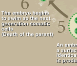
| The volvox is a beautiful, green algae that curls and twines in the water. The newly created body (embryo) reflexes its spherical form and turns completely inside out. Through what mechanism does this phenomenon, known as inversion, occur? The volvox is an interesting model for studying the basics of the creation of form in animals. |
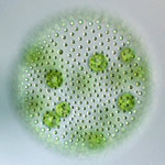 |
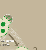
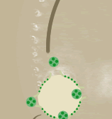
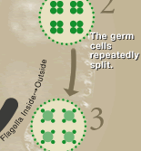
Structure of the volvox body The volvox (V.carteri) used in the experiment were spheroid in shape and 0.5 mm in diameter. About 2,000 small cells (somatic cells) were arrayed in a layer on the surface of the sphere. On the inner surface there are 16 large cells (reproductive cells). The movement of the two flagella on the somatic cells is coordinated, and they paddle the water allowing the volvox to swim. |
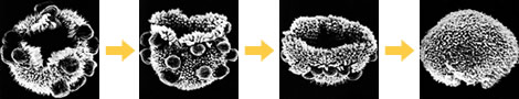 |
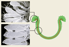
| This is the inversion process shown through electron microscope photography. (The process is shown in Fig.) (Photograph by Dr. David Kirk) |
|
||||
|
| The inversion mechanism |
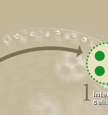
| In the embryos of animals created by the division of zygotes, the neighboring cells are linked in a sheet shape, and this is bent and folded to create the form. The VolvoxÕs form is also created by the sheet, and there is a single layer of cells, making the embryo a simple model. In addition, the germ cells are created before inversion, providing the benefit that mutants with abnormalities in form can be obtained and their genetic background studied. |
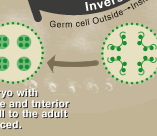
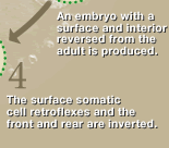
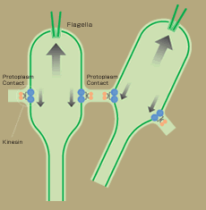
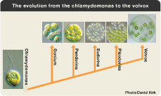
| In the inversion of the volvox embryo, the cell sheet inverts from a cross-shaped hole, and finally the surface and interior are reversed. When the inversion occurs, the cells change shape, becoming long and thin, and the movement of the plasmodesma linking the neighboring cells becomes coordinated. Research using the mutations in which the inversion process has been stopped before completion has presented a model in which the kinecin, the molecular motor created by the gene InvA, moves in one direction in the microtubule while combining with the adhesion strands between the cells and pushes up the cells relative to each other. Perhaps the action of this genetic InvA is the key to multicellularization from the chlamydomonas to the volvox. |
|
||
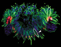
| Left: This is microscope photograph of an embryo during inversion stained by an antibody. The red stained portion is the kinesin. Corresponding with the inversion, the kinesin position moves in relation to that of the green portion (microtube). Center: Electron microscope photographs of cells before and after inversion. In the inverted portion, part of the cell becomes elongated and changes into a bottle shape. Right: A moving model of inversion based on considerations of kinesin movement and changes in the cell shape. |
|
|
|
||
|
|
The reason the freshwater fish arowana live across the sea | ||
| Rsearch |
|
Please close a window with the button of a browser who are turning off Javascript. |