Biohistory Journal, Summer, 2005
Research: Index > Mechanism of symbiosis -Gene network made up of plants and soil microbes
Research
To capture the scene of autophagy
The reason cells eat themselves
Noboru Mizushima,
Tokyo Metropolitan Organization for Medical Research / The Tokyo Metropolitan Institute of Medical Science
    We observed molecules in an attempt to understand metabolism, the invisible flow of energy and substances that support the phenomenon of life, in a visible form. We approached the reason cells eat themselves with a method that dynamically apprehends the form of the living cell. (Text: Editorial staff)

Each point of light is an autophagosome within a fibroblastic cell of a mouse.
The rear cover shows the creation of an autophagosome.
    The cell is the basic unit of organisms. Every day they create the elements required for life and appropriately dispose of them. To use an analogy with human society, this is just as important as creating goods, recycling resources, and disposing of waste material.
    The eukaryote cell has two large-scale degradation systems. The first is the ubiquitin-proteasome system, which recognizes with great precision only the unnecessary proteins and decomposes them. The other is autophagy, which we will discuss here.
    The autophagosome is a structure that surrounds not only proteins but also mitochondria and other organelles and transports them to the lysosome where they are decomposed in a non-selective degradation system. We applied green fluorescent proteins as a marker to the proteins related to autophagy and observed the actual process with a fluorescence microscope.

An enlargement
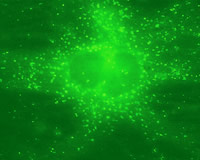
    When, where, and how often does autophagy occur within a body? To find out, we linked a protein (LC3) attached to the autophagosome membrane with the gene of the green fluorescent protein and created a genetically modified mouse to express this throughout its entire body. After 24 hours without food, autophagy was active in most organs. It was extremely interesting that autophagy was temporarily active in different organs in a newborn immediately after birth. In mice, for example, the peak was reached from three to six hours after birth and returned to a low level within a day or two. Newborns in which the supply of nutrition from the umbilical cord was suddenly interrupted were plunged into a severe state of starvation.
    When we produced mice that do not cause autophagy, they had a normal birth, but immediately afterward suffered from severe nutritional deficiency and lowered energy. The role of autophagy is the inevitable decomposition of part of oneself during starvation, and obtaining and recycling the nutritional elements obtained from that process.
    Autophagy also gradually occurs even when the body is not in a state of starvation. This is believed to serve the function as a cleaner inside the cell. In other words, autophagy is thought to have two important roles: to supply nutrition during starvation and to regularly purify the cell interior. The function of autophagy was an enigma for a long time, but it is now rapidly being cleared up.

Masataka Okabe
    Institute of DNA Medicine, Jikei University School of Medicine
    Okabe was awarded a doctorate from the Jikei University School of Medicine in Tokyo in 1996. After serving as a researcher at the Japan Science and Technology Agency (CREST), an assistant professor at the National Institute of Genetics, and a research fellow at King’s College London, he was appointed the head of the Organogenesis Laboratory at the Jikei University School of Medicine’s Institute of DNA Medicine.
 
Moving to land and recycling organs
A scroll - Viewing time
Rsearch

CLOSE

Please close a window with the button of a browser who are turning off Javascript.