論文
Kazuhide Miyamoto, Junpei Kuroda, Satomi Kamimura, Yasuyuki Sasano, Gembu Abe, Satoshi Ansai, Noriko Funayama, Masahiro Uesaka, Koji Tamura
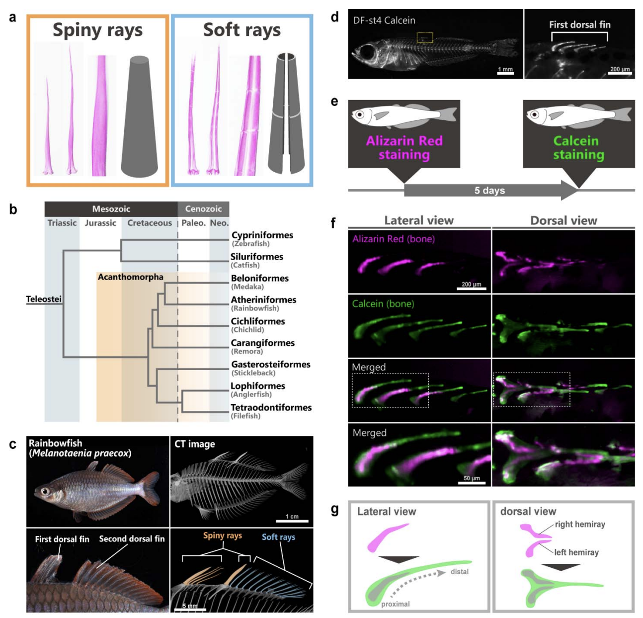
Actinotrichia-independent developmental mechanisms of spiny rays facilitate the morphological diversification of Acanthomorpha fish fins
bioRxiv (2025)
DOI: 10.1101/2025.03.01.640274
解説
Skeletal forms in vertebrates have been regarded as good models of morphological diversification. Fish fin forms are greatly diversified, and their bone structure is classified into soft rays and spiny rays. In fish evolution, spiny-ray morphologies are known to be sometimes extremely modified; however, it remains unknown how the developmental mechanisms of spiny-rays have contributed to their morphological diversification. By using the rainbowfish Melanotaenia praecox for examination of the extracellular matrix (ECM) and cell dynamics of spiny-ray development, we demonstrate that spiny-ray developments are independent of the actinotrichia (needle-shaped collagen polymers at the tip of fish fins), which are known as an important ECM in soft-ray morphogenesis. Furthermore, we found that in the thorny spiny-ray of the filefish Stephanolepis cirrhifer, the lateral protrusions equip the BMP positive osteoblast condensation, as in the state of the spiny-ray tips in M. praecox and S. cirrhifer. Taken together, our findings reveal that osteoblast distribution and signaling-molecule intensity would contribute to spiny-ray modification. In comparison to soft rays development, the independence from actinotrichia in spiny-rays would facilitate growth direction change, leading to their morphological diversification. This indicates that the cell distribution and ECM usage would be major factors driving the morphological diversification in animals.